
Fungal staining is a laboratory technique in microbiology to detect and identify fungi isolated from food or others samples.These methods make transparent fungal structures visible under a microscope,like hyphae, conidia, and spores. There are several staining techniques vary based on the sample type and purpose.We followed Lactophenol Cotton Blue(LPCB) staining procedure in our lab.
Here we have isolated the fungi from Dry Fish Sample.
Chemicals and Equipments Used during Fungal Staining Process:
Chemical Used
- Lactophenol Cotton Blue (LPCB): LPCB (Lactophenol Cotton Blue) is the most widely used mounting and staining medium for visualizing and identifying fungi.
Equipments Used
- Slides : Used to prepare the fungal stain.
- Inoculating needle: Used to safely pick up and transfer fungal cultures to the slide.
- Spirit Lamp : Used to sterilize the inoculating needle.
- Compound Microscope : Used to examine fungi under high magnification.(45x)
- Tissue : It is used for clean the lens and slides.
- Gloves,Cap & Mask : To maintain proper sterility inside the biosafety cabinet. If you don’t wear gloves & masks can affects on analyzer’s body also.
- Cover slips: Small, thin squares of glass placed over the sample to protect the microscope lens.
- Bio Safety Cabinet: Used to conduct the process in a sterile area for safety.
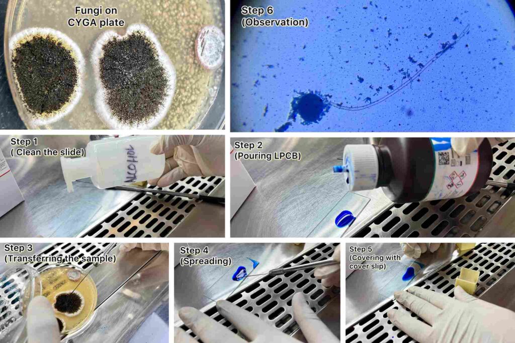
Steps followed in Fungal Staining process:
Prepare a Slide: Place a drop of Lactophenol Cotton Blue stain on a clean glass slide.
│
▼
Transfer the Sample: Gently take a small portion of the fungal culture using a sterile inoculating needle.
│
▼
Spread the Sample: Place the fungus into the drop of stain and use needle to spread it thinly.
│
▼
Add Coverslip: Carefully place a coverslip at an angle to avoid trapping air bubbles.
│
▼
Examine: Observe under a microscope using low (10x) to high (45x) power.
Why Fungal staining is needed to do?
- To study the morphology & structural differences in fungal cell .
- To help in the preliminary identification of fungi .
- To observe fungal shape, arrangement, and staining characteristics under a microscope.
- To identify the different strain of fungi.
Basic Handling Mistakes to be avoided during Fungal staining:
- Ensure the slide is clean & grease-free for clear observation.Use clean glasswares.Washing procedure described.
- Adding too much stain washes out the specimen.Apply exactly one to two drops of stain.
- Carefully place a coverslip at an angle to avoid trapping air bubbles.
- Work inside a biological safety cabinet (BSC) for safety.
Conclusion Of Fungal Staining Process:
Fungal staining is used to detect and identify fungi .This method is easy & widely followed to observe the fungal structures under the microscope. By following these method,you can easily do the Fungal Staining procedure at any laboratory or manufacturing industry with availability of the equipment & chemicals.If you can’t understand the procedure you can check our real time photo attached with this writing or also you can reach to Pro Research & Testing Laboratory for the testing purposes.
FAQ on Fungal Staining
Q1:What is Fungal staining ?
Ans:Fungal staining is a laboratory technique in microbiology to observe morphology of fungi.
Q2: What mistake may affect the result during Fungal staining ?
Ans:Adding too much stain washes out the specimen.
Q3: Why Grease-free slide use is important in Fungal staining?
Ans:Grease-free slide use is important in Fungal staining for clear observation.Unwanted particles can create problem to view exact structure.
Q4: Why Spirit lamp is use in Fungal staining?
Ans:It is Used to sterilize the inoculating needle.
Q5:What is the purpose of the Fungal staining procedure?
Ans: To help in the preliminary identification of Fungi .
How We Verified This Testing/Research Procedure :
This testing procedure is done under qualified analyst .Continually monitored by expertise.Repeatedly testing is always done to get accurate result.

Written by
Ankita Samanta (M.Sc Microbiology,Vidyasagar University)
Designation – Junior Microbiologist
Reviewed by
Anwesha Das (M.Sc Microbiology,BU)
Designation – Microbiologist
Verified By
Tathagata Talukdar (M.Sc,Microbiology) University of Calcutta
Designation – Senior/Chief Microbiologist
Experience – 12 Years + of experience including medical microbiology (NABL 15189) and general microbiology (NABL 17025)