
Gram staining is the differential staining technique in microbiology. It classifies bacteria into two major groups Gram-positive and Gram-negative, based on the thickness of their peptidoglycan cell wall. In the gram staining procedure , we followed four distinct steps that are Primary Stain (Crystal Violet), Mordant (Gram’s Iodine),Decolorization (Acetone) & Counterstain (Safranin).
Here we are doing Gram Staining test of Escherichia Coli bacteria.
Chemicals and Equipments Used during Gram Staining Process:
Chemical Used
- Crystal Violet : Crystal violet is primarily used as the primary stain in Gram staining. It penetrates bacterial cell walls, dyeing them purple.
- Gram’s Iodine : Gram’s iodine serves as a mordant by forming a crystal violet–iodine complex, which helps fix the primary stain inside the cells.
- Acetone : Acetone acts as a decolorizer that removes the primary stain from Gram-negative bacteria.
- Safranin : Safranin is used as a counterstain in the Gram staining procedure,giving decolorized cells a pink or red color.
- Sterile Distilled Water: Sterile distilled water is used for preparing the bacterial smear and for rinsing between different staining steps.
Equipments Used
- Slides : Used to prepare the bacterial smear.
- Inoculation Loop : Used to transfer bacterial colonies safely.
- Spirit Lamp : Required during heat-fixing the bacteria onto the slide.
- Staining Rack : To hold the slide and wash away excess reagents during steps.
- Compound Microscope : Used to examine bacteria under high magnification.
- Immersion oil : Applied on the slide to improve resolution when using the 100x objective lens.
- Tissue : It is used for clean the lens and slides.
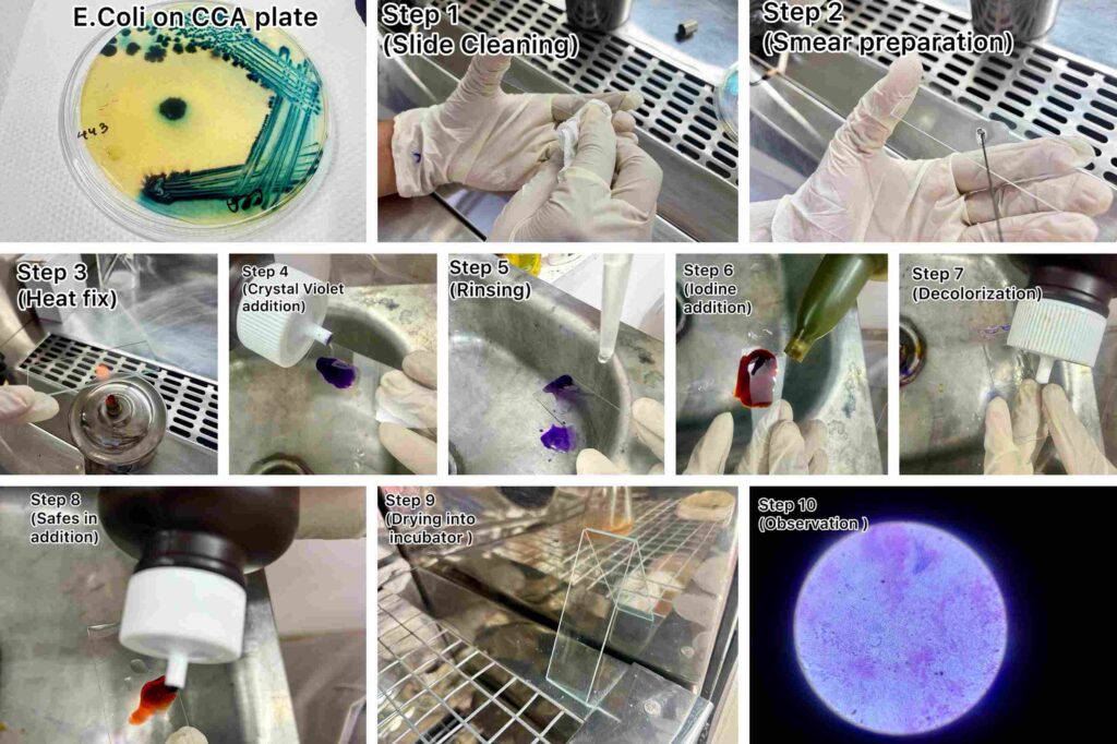
Steps followed in Gram Staining process:
| Steps | Description |
|---|---|
| Prepare the Slide & Fix the smear | A thin smear of the bacterial sample is prepared on a clean glass slide using a sterile loop and a drop of water. The smear is then air-dried and heat-fixed by passing the slide gently through a flame to make the cells stick to the slide. |
| Primary Stain (Crystal violet) | The heat-fixed smear is covered with crystal violet stain and left for about 1 minute. After staining, it is gently rinsed with water to remove excess dye. This step stains all bacterial cells purple initially. |
| Mordant (Gram’s Iodine) | Add iodine solution to fully cover the slide and allow it to react for 2 minutes to form a crystal violet–iodine complex, then gently rinse with water again. |
| Decolorization | Decolorizat is added to the slide for a few seconds to remove excess stain. It is then immediately rinsed with water to stop the reaction |
| Counterstain (Safranin) | Safranin stain is added to the slide and left for about 1 minute to stain the decolorized cells, then gently rinsed with water. This provides contrast, allowing Gram-negative bacteria to appear pink/red while Gram-positive bacteria remain purple. |
| Dry & Observation | Dry the slide inside the incubator and observe the bacterial cell under microscope using 45x & 100x oil immersion objective.Study the shape, arrangement, and staining characteristics of the bacteria and recorded as either Gram-positive (purple) or Gram-negative (pink/red). |
| Result | Escherichia coli (E. coli) is observed under a microscope after Gram staining, it appears as Gram-negative bacteria. They stain pink or red due to the uptake of safranin after decolorization. The cells are short rod-shaped (bacilli). |
What is the purpose of the Gram staining procedure?
- To differentiate bacteria into Gram-positive and Gram-negative groups.
- To study the morphology & structural differences in bacterial cell walls.
- To help in the preliminary identification of bacteria.
- To observe bacterial shape, arrangement, and staining characteristics under a microscope.
Care should be taken to avoid handling mistakes during Gram staining:
- Ensure the slide is grease-free for clear observation.
- Prepare a thin smear using a small inoculum and heat-fix it properly.
- Let the smear air dry completely at room temperature before heat-fixing.
- Pass the slide through the spirit lamp flame only 2–3 times during heat-fixation.
- Do not over-decolorize or under-decolorize with acetone.
- Rinse each stain gently to avoid washing away the smear.
- Avoid using contaminated glasswares.Proper cleaning Process described previously.
- Follow correct timing for each staining step.
Conclusion Of Gram Staining Process:
Gram staining is a technique used to quickly classify bacteria into two categories Gram-positive and Gram-negative.Its primary purpose is to quickly identify bacteria based on it’s morphology. Gram Positive bacteria appear deep purple under a microscope & Gram Negative appear deep pink under a microscope.By following these method,you can easily do the Gram Staining procedure at any laboratory or manufacturing industry with availability of the equipment & chemicals.If you can’t understand the procedure you can check our real time photo attached with this writing or also you can reach to Pro Research & Testing Laboratory for the testing purposes.
FAQ on Gram Staining
Q. What is Gram staining ?
A. Gram staining is a vital microbiological technique used to rapidly identify and classify bacteria.
Q. How many steps are there in Gram staining ?
A. There are four distinct steps in Gram staining.1)Primary Stain (Crystal Violet), 2)Mordant (Gram’s Iodine),3)Decolorization (Acetone) & 4)Counterstain (Safranin).
Q. What mistake may affect the result during Gram staining ?
A. Prepare a thin smear using a small inoculum and heat-fix it properly.A thick smear leads to uneven staining and makes it difficult to clearly observe and correctly identify bacteria under the microscope.
Q. Why Grease-free slide use is important in Gram staining?
A. A grease-free slide is important in Gram staining because any oil or grease can prevent the smear from spreading evenly and stop proper adhesion of bacteria to the slide.
Q. Why decolorizer is used in Gram staining ?
A. Without the decolorization step, all bacteria would remain purple from the initial crystal violet stain.
How We Verified This Testing/Research Procedure :
This testing procedure is done under qualified analyst .Continually monitored by expertise.Repeatedly testing is always done to get accurate result.

Written by
Ankita Samanta (M.Sc Microbiology,Vidyasagar University)
Designation – Junior Microbiologist
Reviewed by
Anwesha Das (M.Sc Microbiology,BU)
Designation – Microbiologist
Verified By
Tathagata Talukdar (M.Sc,Microbiology) University of Calcutta
Designation – Senior/Chief Microbiologist
Experience – 12 Years + of experience including medical microbiology (NABL 15189) and general microbiology (NABL 17025)